 |
|
|
|
Sun, 26 Jul, 2026
|

|

|
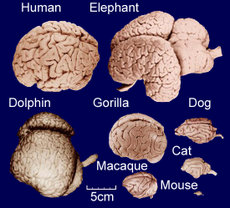 Comparative brain sizes
In animals, the brain, or encephalon ( Greek for "in the head"), acts as the control center of the central nervous system. In most animals, the brain is located in the head close to the primary sensory apparatus and the mouth. While all vertebrate nervous systems have a brain, invertebrate nervous systems may have either a centralized brain or collections of individual ganglia. The brain is an extremely complex organ; for example, the human brain is a collection of 100 billion neurons, each linked with up to 25,000 others [1]. This huge number of interconnecting neurons—often referred to as a neural ensemble—is what allows the brain to conduct such complex processes.The brain controls and coordinates most sensory systems, movement, behaviour, and homeostatic body functions such as heart rate, blood pressure, fluid balance, and body temperature. The brain is the source of cognition, emotion, memory, and motor, and other forms of learning. Many behaviors such as simple reflexes and basic locomotion, can be executed under spinal cord control alone.Most brains exhibit a visible distinction between grey matter and white matter. Grey matter consists of the cell bodies of the neurons, while the white matter consists of the fibers ( axons) that connect neurons over long distances. The entire outer visible layers of the brain is called the cortex which consists primarily of grey matter. However, deeper grey matter structures called nuclei also exist throughout the central nervous system. The axons of this white matter are surrounded by a fatty insulating sheath called myelin, giving the white matter its distinctive colour.The study of the brain is known as neuroscience, a field of biology aimed at understanding the functions of the brain at every level, from the molecular up to the psychological.
 Jump to Page Contents Jump to Page Contents
|
|

Pay as you go
No monthly charges. Access for the price of a phone call
Go>
Unmetered
Flat rate dialup access from only £4.99 a month Go>
Broadband
Surf faster from just £13.99 a month Go> |
Save Even More
Combine your phone and internet, and save on your phone calls
More Info> |
This weeks hot offer
 24: Series 5
In association with Amazon.co.uk £26.97 |
|
Contents
History
Modern neuroscience
Mind and brain
Comparative anatomy
Neurobiology
The study of the brain
Brain as food
History - Contents
 Hieroglyphic for the word "brain" (c.1700 BC)
The Edwin Smith Surgical Papyrus, written in the 17th century BC, contains the earliest reference to the brain anywhere in human records. The word brain (adjacent), occuring only eight times in this papyrus, describes the symptoms, diagnosis, and prognosis of two patients, wounded in the head, who had compound fractures of the skull.In the first millennium BC, the Ancient Greeks developed differing views on the function of the brain. In the 4th BC Hippocrates, the father of medicine, believed the brain to be the seat of intelligence. In the 3rd century BC Aristotle believed that the brain was a cooling mechanism for the blood while the heart was the seat of intelligence. He reasoned that humans are more rational than the beasts because they have a proportionally larger brain to cool their hot-bloodedness .
 Drawing of the base of the brain by Andrea Vesalius (1543).
During the Roman Empire, the anatomist Galen dissected the brains of sheep. He concluded that since the cerebellum was more dense than the brain, it must control the muscles. Since cerebrum was soft, it must be where the senses were processed. Galen further theorized that the brain functioned by movement of fluids through the ventricles.In the Age of Enlightenment, René Descartes espoused a fluid mechanical view of the brain similar to Galen's theories. However, Descartes thought that although this explanation was adequate to explain the brain functions of animals, the higher mental functions of humans were accomplished by the soul. This theoretical separation of the mind and brain became known as the mind-body problem, with Descartes espousing the dualist view of the mind existing separate from the brain.
 Drawing of the cells in the chicken cerebellum by S. Ramón y Cajal
In the mid- 1600s, however, great progress in describing the anatomy of the brain ( neuroanatomy) was achieved with the works of English anatomist Thomas Willis and Flemish anatomist Vesalius. They dispelled many of the notions of Galen and Descartes, and resolved many facts about the macro structure of the brain of animals and humans.In the 1700s, Luigi Galvani showed that electrically stimulating the sciatic nerve of a dissected frog caused movement of the attached muscle. His experiments moved scientists away from the fluid mechanical theory of the brain and toward an electrical theory. In the 19th century, Galvani's work led to the development of research in bioelectricity and to the discovery of the membrane potential and action potential by researchers such as Emil du Bois-Reymond.The scientists of the 1800s debated whether areas of the brain corresponded to specific functions or if the brain functioned as a whole (the "aggregate field theory"). Jean Pierre Flourens championed the aggregate field theory in opposition to the pseudoscience of phrenology, founded by Franz Joseph Gall. However, the work of Paul Pierre Broca, Karl Wernicke, and Korbinian Brodmann eventually helped to show that areas of the brain had specific functions. Their work showed that, while some functions were repeated, there is also a lateralization of brain function wherein certain functions such as speech and language are usually controlled by one cerebral hemisphere as opposed to another. The redundancy of functioning has come to be known as parallel distributed processing .
 First EEG trace recorded by Hans Berger (1929)
By the 20th century, the anatomical works of Santiago Ramón y Cajal and Camillo Golgi laid the foundation for the study of individual neurons in the brain. Charles Scott Sherrington and Edgar Douglas Adrian furthered the study of neurons with the new techniques using electrodes. Neurotransmitters were discovered and investigated by a number of scientists, including Otto Loewi, Henry Hallett Dale, Arvid Carlsson, and many others. These neurochemicals are responsible for carrying signals from one neuron to another across the tiny gaps ( synapses) that exist between the neuronal connections.In 1929, German physician Hans Berger recorded the first electrical potentials from a living brain. This technique—known as electroencephalography or EEG—led to the widespread use of neuroimaging on live, active humans and animals to study the processes of the mind.
Modern neuroscience - Contents
Modern neuroscience is experiencing rapid growth due to the availability of computers capable of handling the intense processing required for understanding such a complex system. Neuroscientists use a variety of approaches to study the brain at different levels—from the molecules to systems. Extensive knowledge has been accumulated about the electrophysiological properties of different types of neurons and their responsiveness to neurotransmitters. Recordings from the brains of awake, behaving animals pioneered by Edward Evarts help to decode neuronal firing during different behaviors and cognitive processes. Miguel Nicolelis introduced multielectrode recording techniques which led to creation of rudimentary brain-computer interfaces. Rapidly developing neuroimaging techniques such as functional magnetic resonance imaging (fMRI) allows scientists to study the brain in living humans and animals in ways that their predecessors could not.
Mind and brain - Contents
A distinction is sometimes made in the philosophy of mind between the mind and brain. The brain is defined as the physical, biological matter contained within the head, responsible for all electrochemical neuronal processes. The mind, however, exists as something outside of the brain. The mind is sometimes thought of as consciousness, the soul, or some other non-physical center of thought.The inability to determine what consciousness is has led to the mind-body problem. Some philosophers such as strong AI theorists believe that the mind is analogous to computer software and the brain to hardware.
Comparative anatomy - Contents
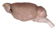 A mouse brain.
Three groups of animals, with some exceptions, have notably complex brains: the arthropods (insects and crustaceans), the cephalopods (octopuses, squid, and similar mollusks), and the craniates ( vertebrates). The brain of arthropods and cephalopods arises from twin parallel nerve cords that extend through the body of the animal. In arthropods, the brain consists of a central brain with three divisions and large optical lobes behind each eye for visual processing.The brain of craniates develops from the anterior section of a single dorsal nerve cord, which later becomes the spinal cord. In craniates, the brain is protected by the bones of the skull. In vertebrates, increasing complexity in the cerebral cortex correlates with height on the phylogenetic and evolutionary tree. Primitive vertebrates such as fish, reptiles, and amphibians have fewer than six layers of neurons in the outer layer of their brains. This cortical configuration is called the allocortex (or heterotypic cortex).More complex vertebrates such as mammals have developed a six-layered neocortex (or homotypic cortex, neopallium), in addition to having some parts of the brain that are allocortex. In mammals, increasing convolutions of the brain are characteristic of animals with more advanced brains. These convolutions evolved to provide a larger surface area for a greater number of neurons while keeping the volume of the brain compact enough to fit inside the skull. This folding allows for more grey matter to fit into a smaller volume, similar to a really long slinky being able to fit into a tiny box when completely pushed together. The folds of the brain are called gyri, while the spaces between the folds are called the sulci.Although the general histology of the brain is common to all those who have one, the structural anatomy differs from person to person. Apart from the gross embryological divisions of the brain, the individual location of specific gyri and sulci, primary sensory regions, and other structures in relation to one another differs across creatures.
Invertebrates
In insects, the brain can be divided into four parts, the optical lobes, the protocerebrum, the deutocerebrum, and the tritocerebrum. The optical lobes are positioned behind each eye and process visual stimuli . The protocerebrum contains the mushroom bodies, which respond to smell, and the central body complex. In some species such as bees, the mushroom body receives input from the visual pathway as well. The deutocerebrum includes the antennal lobes, which are similar to the mammalian olfactory bulb, and the mechanosensory neuropils which receive information from touch receptors on the head and antennae. The antennal lobes of flies and moths are quite complex.In cephalopods, the brain is divided into two regions: the supraesophageal mass and the subesophageal mass . These parts are divided by the animal's esophagus. The supra- and subesophageal masses are connected to each other on either side of the esophagus by the basal lobes and the dorsal magnocellular lobes . The large optic lobes are sometimes not considered to be part of the brain proper since the optic lobes are anatomically separate from the brain and are joined to the brain by the optic stalks. However, the optic lobes perform much of the visual processing and functionally can be considered part of the brain.
Vertebrates
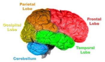 The lobes of the cerebral cortex include the frontal (red), temporal (green), occipital (yellow), and parietal lobes (orange). The cerebellum (blue) is not part of the telencephalon. In vertebrates a gross division into three major parts is used.
The telencephalon (cerebrum) makes up the largest section of the mammalian brain. This is the structure that is most easily visible, and is what most people associate with the "brain". In humans the fissures (sulci) and convolutions (gyri) give the brain a wrinkled appearance. In most vertebrates the metencephalon is the highest center in the brain, whereas in mammals this role has been adopted by the cerebrum. Because humans walk upright, this creates a flexure, or bent nature, to the brain between the brain stem and the cerebrum that is not present in most other vertebrates. For this reason, descriptions of the locations of certain brain structures in humans as compared to other vertebrate species may be confusing.Behind (or in humans, below) the cerebrum is the cerebellum. The cerebellum functions mainly in the control of movement and movement timing . It is connected via thick white matter fibers (cerebellar peduncles) to the pons. The cerebrum and the cerebellum each consist of two hemispheres. The telencephalic hemispheres are connected by the corpus callosum, another large white matter tract. An outgrowth of the telencephalon called the olfactory bulb is a major structure in many animals, but in humans and other primates it is relatively small.Vertebrate nervous systems are distinguished by encephalization and bilateral symmetry. Encephalization refers to the tendency for more complex organisms to gain larger brains through evolutionary time. Larger vertebrates develop a complex system of layered and interconnected neuronal circuitry. In modern species most closely related to the first vertebrates, brains are covered with gray matter that has a three-layer structure (allocortex). Their brains also contain deep brain nuclei and fiber tracts forming the white matter. Most regions of the human cerebral cortex have six layers of neurons (neocortex) .Vertebrate brain regions
(See related article at List of regions in the human brain)
 Diagram depicting the main subdivisions of the embryonic vertebrate brain. These regions will later differentiate into forebrain, midbrain and hindbrain structures.
According to the hierarchy based on embryonic and evolutionary development, chordate brains are composed of the three regions that later develop into five total divisions:
-
Rhombencephalon (hindbrain)
-
Myelencephalon
-
Metencephalon
-
Mesencephalon (midbrain)
-
Prosencephalon (forebrain)
-
Diencephalon
-
Telencephalon
The brain can also be classified according to function, including divisions such as:
-
Limbic system
-
Sensory systems
-
Visual system
-
Olfactory system
-
Gustatory system
-
Auditory system
-
Somatosensory system
- Motor system
-
Associative areas
Humans
The structure of the human brain is different from that of other animals in several significant ways. These differences have allowed for many abilities over and above those of other animals, such as advanced cognitive skills. Human encephalization is especially pronounced in the neocortex, the most complex part of the cerebral cortex. The proportion of the human brain that is devoted to the neocortex—especially to the prefrontal cortex—is larger than in all other animals.Humans enjoy unique neural capacities, but much of the human brain structure is shared with ancient species. Basic systems that alert the nervous system to stimulus, that sense events in the environment, and monitor the condition of the body are similar to those of the most basic vertebrates. The neural circuitry underlying human consciousness includes both the advanced neocortex and prototypical structures of the brain stem. The human brain also has a massive number of synaptic connections allowing for a great deal of parallel processing.
Neurobiology - Contents
Despite the variance of the species in which the brain is found there are many common features in its cellular make-up, its structure, and its function. On a cellular level the brain is composed of two classes of cells, neurons and glia, both of which contain several different cell types which perform different functions. Interconnected neurons form neural networks (or neural ensembles). These networks are similar to man-made electrical circuits in that they contain circuit elements (neurons) connected by biological wires (nerve fibers). These do not form simple one-to-one electrical circuits like many man-made circuits, however. Typically neurons connect to at least a thousand other neurons. These highly specialized circuits make up systems which are the basis of perception, action, and higher cognitive function.
Histology
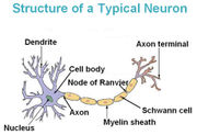 Diagram of basic features of a neuron.
Neurons are the cells that generate action potentials and convey information to other cells; these constitute the essential class of brain cells. In each particular brain area, input (or afferent) neurons, output (or efferent) neurons, and interneurons are typically found. Input neurons are recipients of projections from other brain areas. Output neurons project to the other areas. Interneurons are the neurons which do not leave the area but rather perform local processing.In addition to neurons, the brain contains glial cells in a roughly 10:1 proportion to neurons. Glial cells (Greek: “glue”) perform supportive roles to neurons including creating the insulating myelin, providing structure to the neuronal network, waste management, and neurotransmitter clean up. Most types of glia in the brain (and the rest of the central nervous system) are present in the entire nervous system. Exceptions include oligodendrocytes which insulate neural axons (a role performed by Schwann cells in the peripheral nervous system). Oligosaccharides are the defining factor between white matter and grey matter in the brain—white matter is composed of myelinated axons, whereas grey matter contains mostly cell soma, dendrites, and unmyelinated portions of axons and glia. The space between neurons is filled with dendrites as well as unmyelinated segments of axons; this area is referred to as the neuropil.In mammals, the brain also contains connective tissue called the meninges, a system of membranes that separate the skull from the brain. This three-layered covering is made of, from the outside in, dura mater, arachnoid mater, and pia mater. The arachnoid and pia are physically connected and thus often considered as a single layer, the pia-arachnoid. Below the arachnoid is the subarachnoid space which contains cerebrospinal fluid, a substance that protects the nervous system. Blood vessels enter the central nervous system through the perivascular space above the pia mater. A blood-brain barrier protects the brain from toxins that might enter through the blood.The brain is suspended in cerebrospinal fluid (CSF), which circulates between layers of the meninges and through cavities in the brain called ventricles. It is important both chemically for metabolism and mechanically for shock-prevention. For example, the human brain weighs about 1-1.5 kg. The mass and density of the brain are such that it will begin to collapse under its own weight. The CSF allows the brain to float, easing the stress caused by the brain’s mass.
Function
Vertebrate brains receive signals through nerves arriving from the sensors of the organism. These signals are then interpreted throughout the central nervous system reactions are formulated based upon reflex and learned experiences. A similarly extensive nerve network delivers signals from a brain to control muscles throughout the body. Anatomically, the majority of afferent and efferent nerves (with the exception of the cranial nerves) are connected to the spinal cord, which then transfers the signals to and from the brain.Sensory input is processed by the brain to recognize danger, find food, identify potential mates, and perform more sophisticated functions. Visual, touch, and auditory sensory pathways of vertebrates are routed to specific nuclei of the thalamus and then to regions of the cerebral cortex that are specific to each sensory system. The visual system, the auditory system, and the somatosensory system. Olfactory pathways are routed to the olfactory bulb, then to various parts of the olfactory system. Taste is routed through the brainstem and then to other portions of the gustatory system.To control movement the brain has several parallel systems of muscle control. The motor system controls voluntary muscle movement, aided by the motor cortex, cerebellum, and the basal ganglia. The system eventually projects to the spinal cord and then out to the muscle effectors. Nuclei in the brain stem control many involuntary muscle functions such as heart rate and breathing. In addition, many automatic acts (simple reflexes, locomotion) can be controlled by the spinal cord alone.Brains also produce a portion of the body's hormones that can influence organs and glands elsewhere in a body—conversely, brains also react to hormones produced elsewhere in the body. In mammals, most of these hormones are released into the circulatory system by a structure called the pituitary gland.It is hypothesized that developed brains derive consciousness from the complex interactions between the numerous systems within the brain. Cognitive processing in mammals occurs in the cerebral cortex but relies on midbrain and limbic functions as well. Among "younger" (in an evolutionary sense) vertebrates, advanced processing involves progressively rostral (forward) regions of the brain.Hormones, incoming sensory information, and cognitive processing performed by the brain determine the brain state. Stimulus from any source can trigger a general arousal process that focuses cortical operations to processing of the new information. This focusing of cognition is known as attention. Cognitive priorities are constantly shifted by a variety of factors such as hunger, fatigue, belief, unfamiliar information, or threat. The simplest dichotomy related to the processing of threats is the fight-or-flight response mediated by the amygdala and other limbic structures.
Brain pathology
 A human brain showing frontotemporal lobar degeneration causing frontotemporal dementia.
Clinically, death is defined as an absence of brain activity as measured by EEG. Injuries to the brain tend to affect large areas of the organ, sometimes causing major deficits in intelligence, memory, and movement. Head trauma caused, for example, by vehicle and industrial accidents, is a leading cause of death in youth and middle age. In many cases, more damage is caused by resultant swelling (edema) than by the impact itself. Stroke, caused by the blockage or rupturing of blood vessels in the brain, is another major cause of death from brain damage.Other problems in the brain can be more accurately classified as diseases rather than injuries. Neurodegenerative diseases, such as Alzheimer's disease, Parkinson's disease, motor neurone disease, and Huntington's disease are caused by the gradual death of individual neurons, leading to decrements in movement control, memory, and cognition. Currently only the symptoms of these diseases can be treated. Mental illnesses, such as clinical depression, schizophrenia, bipolar disorder, and post-traumatic stress disorder are brain diseases that impact the personality and typically on other aspects of mental and somatic function. These disorders may be treated by psychiatric therapy, pharmaceutical intervention, or through a combination of treatments; therapeutic effectiveness varies significantly among individuals.Some infectious diseases affecting the brain are caused by viral and bacterial infection(s). Infection of the meninges, the membrane that covers the brain, can lead to meningitis. Bovine spongiform encephalopathy (also known as mad cow disease), is deadly in cattle and is linked to prions. Kuru is a similar prion-borne degenerative brain disease affecting humans. Both are linked to the ingestion of neural tissue, and may be an evolutionary defense against cannibalism. Viral or bacterial causes have been substantiated in multiple sclerosis, Parkinson's disease, Lyme disease, encephalopathy, and encephalomyelitis.Some brain disorders are congenital. Tay-Sachs disease, Fragile X syndrome, and Down syndrome are all linked to genetic and chromosomal errors. Malfunctions in the embryonic development of the brain can be caused by genetic factors, by drug use, and disease during a mother's pregnancy.
The study of the brain - Contents
Fields of study
Several areas of science specifically study the brain. Neuroscience seeks to understand the nervous system, including the brain, from a biological and computational perspective. Psychology seeks to understand behaviour and the brain. The terms neurology and psychiatry usually refer to medical applications of neuroscience and psychology respectively. Cognitive science seeks to unify neuroscience and psychology with other fields that concern themselves with the brain, such as computer science ( artificial intelligence and similar fields) and philosophy.
Methods of observation
Electrophysiology
Each method for observing activity in the brain has its advantages and drawbacks. Electrophysiology, in which wire electrodes are implanted in the brain, allows scientists to record the electrical activity of individual neurons or fields of neurons. However this method requires invasive surgery and thus this technique is typically usually used only with lab animals or during neurosurgery.EEG
By placing electrodes on the scalp one can record the summed electrical activity of the cortex in a technique known as EEG. EEG measures the mass changes in electrical current from the cerebral cortex, but can only detect changes over large areas of the brain with very little sub-cortical activity.fMRI and PET
Functional magnetic resonance imaging (fMRI) measures changes in blood flow in the brain, but the activity of neurons is not directly measured, nor can it be distinguished whether this activity is inhibitory or excitatory. Similarly, a positron emission tomography (PET), is able to monitor glucose metabolism in different areas within the brain which can be correlated to the level of activity in that region.Behavioral
Behavioral tests can measure symptoms of disease and mental performance, but can only provide indirect measurements of brain function and may not be practical in all animals. In humans however, a neurological exam can be done to determine the location of any trauma, lesion, or tumor within the brain, brain stem, or spinal cord.Anatomical
Autopsy analysis of the brain allows for the study of anatomy and protein expression patterns, but is only possible after the human or animal is dead. Magnetic resonance imaging (MRI) can be used to study the anatomy of a living creature and is widely used in both research and medicine.Other methods
Attempts have also been made to directly "read" the brain, which has been accomplished in a rudimentary manner through a brain-computer interface. Brain activity can be detected by implanted electrodes, raising the possibility of direct mind-computer interface. The reverse method has been successfully demonstrated: brain implants have been used to generate artificial hearing and (crude and experimental) artificial vision for deaf and blind people. Brain pacemakers are now commonly used to regulate brain activity in conditions such as Parkinson's disease.
Other matters
Computer scientists have produced simulated neural networks loosely based on the structure of neuron connections in the brain. Artificial intelligence seeks to replicate brain function—although not necessarily brain mechanisms—but as yet has been met with limited success.Creating algorithms to mimic a biological brain is extremely difficult because the brain is not a static arrangement of circuits, but rather a network of vastly interconnected neurons that are constantly changing their connectivity and sensitivity. More recent work in both neuroscience and artificial intelligence models the brain using the mathematical tools of chaos theory and dynamical systems. Current research has also focused on recreating the neural structure of the brain with the aim of producing human-like cognition and artificial intelligence.
Brain as food - Contents
Like most other internal organs, the brain can serve as nourishment. For example, in the Southern United States canned pork brain in gravy can be purchased for consumption as food. The form of brain is often fried with scrambled eggs to produce the famous "Eggs n' Brains".The brain of animals also features in French cuisine such as in the dish [tête de veau], or head of calf. Although it might consist only of the outer meat of the skull and jaw, the full meal includes the brain, tongue, and glands, with the latter form being the favorite food of French President Jacques Chirac.Similar delicacies from around the world include Mexican tacos de sesos made with cattle brain as well as squirrel brain in the US South. The Anyang tribe of Cameroon practiced a tradition in which a new tribal chief would consume the brain of a hunted gorilla while another senior member of the tribe would eat the heart.Consuming the brain and other nerve tissue of animals is not without its risks. The first problem is that the brain is made up of 60% fat due to the myelin (which by itself is 70% fat) insulating the axons of neurons and glia. As an example, a 0.14 kg can of "pork brains in milk gravy", a single serving, contains 3500 milligrams of cholesterol, 1170% of our recommended daily intake. Brain consumption can also result in contracting fatal transmissible spongiform encephalopathies such as Creutzfeldt-Jakob disease and other prion diseases in humans and mad cow disease in cattle.Another prion disease called kuru has been traced to a mourning ritual among the Fore people of Papua New Guinea in which those close to the dead would eat their brain to create a sense of immortality. Some archaeological evidence suggests that the mourning rituals of European Neanderthals also involved the consumption of the brain.The practice of eating another human's brain has been depicted by Hollywood in the film Hannibal and countless zombie movies.It is not only humans who eat the brains of other animals. The two species of chimpanzee, though generally vegetarian, are known to eat the brains of monkeys to obtain fat in their diet. |
Change Text Size:
[A]
[default]
[A] |


 |
|
|
|
|